Leg Bones Diagram - Human Leg Bones / They allow you to move and provide support for your upper body.. At the microscopic level, this hard outer. Bone surfaces at synovial joints are protected by a coating of articular cartilage. However, the definition in human anatomy refers only to the section of the lower limb extending from the knee to. Bones of the leg and foot, lower leg bone anatomy, leg bones anatomy, leg muscles, leg bones diagram, leg bone structure, leg anatomy muscles, parts of the lower leg. Your legs are two of your most important body parts.
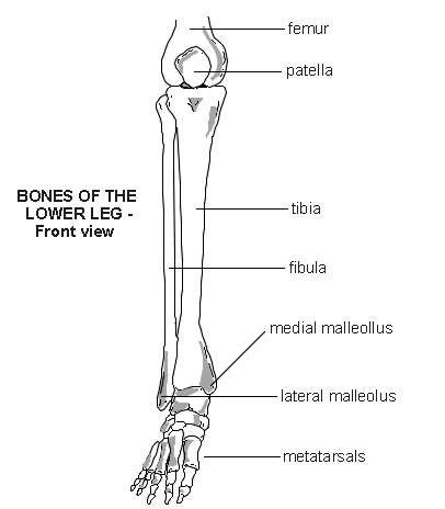
Quizzes on human skeletal system anatomy, bone anatomy, and bone markings. Interactive anatomical atlas of the head, brain, and neck based on anatomical diagrams and ct and mri medical imaging exams. Master leg and knee anatomy using our topic page. Bone surfaces at synovial joints are protected by a coating of articular cartilage. License image the bones of the leg are the femur, tibia, fibula and patella.
Lower leg - bones | Diagram | Patient from patient.azureedge.net Master leg and knee anatomy using our topic page. Your legs are two of your most important body parts. The foot bones shown in this diagram are the talus, navicular, cuneiform, cuboid, metatarsals and calcaneus. Pngtree offers bone diagram png and vector images, as well as transparant background bone diagram clipart images and psd files. Your leg bones are the longest and strongest bones in your body. Describe the bones and bony landmarks that articulate at each joint of the lower limb. Human foot bones anatomy sketch of orthopedics medicine. Synovial joints are often supported and reinforced by surrounding ligaments, which limit movement to prevent injury.
This long bone connects with the knee at one end and the next to the tibia is the fibula, the thinner, weaker bone of the lower leg.
Describe the bones and bony landmarks that articulate at each joint of the lower limb. Cheek bone (zygoma) upper jaw (maxilla). The foot bones shown in this diagram are the talus, navicular, cuneiform, cuboid, metatarsals. High quality realistic skeleton legs. You'll learn about the muscles, bones, and other structures of each area of the leg. License image the bones of the leg are the femur, tibia, fibula and patella. When you stand or walk, all the weight of your upper body rests on them. Pngtree offers bone diagram png and vector images, as well as transparant background bone diagram clipart images and psd files. High resolution textures and displacement included. The largest and most medial leg bone, forming both the knee and ankle joints. Time to jump right into the biggest and strongest bones in the human body. Learn vocabulary, terms and more with flashcards, games and other study tools. At the microscopic level, this hard outer.
At the microscopic level, this hard outer. Health diagram bone skeleton leg knee science anchor chart human human body. Pngtree offers bone diagram png and vector images, as well as transparant background bone diagram clipart images and psd files. You'll learn about the muscles, bones, and other structures of each area of the leg. The foot bones shown in this diagram are the talus, navicular, cuneiform, cuboid, metatarsals and calcaneus.
Nerves of the Leg and Foot | Interactive Anatomy Guide from www.innerbody.com Interactive anatomical atlas of the head, brain, and neck based on anatomical diagrams and ct and mri medical imaging exams. Describe the bones and bony landmarks that articulate at each joint of the lower limb. However, the definition in human anatomy refers only to the section of the lower limb extending from the knee to. High quality realistic skeleton legs. Most bones (particularly the long bones of the arms and legs — which make up the appendicular skeleton) have a hard outer shell known as cortical bone. The bones involved in it, however, are only the femur and the tibia, although the smaller bone of the leg, the fibula, is carried along in the movements of flexion, extension, and slight rotation that this joint. Visit kenhub for more skeletal system quizzes. Bone surfaces at synovial joints are protected by a coating of articular cartilage.
They allow you to move and provide support for your upper body.
Most bones (particularly the long bones of the arms and legs — which make up the appendicular skeleton) have a hard outer shell known as cortical bone. Each leg is made up of four bones. The musculoskeletal segment of the leg, including the foot bones (ankle, heel bone, toe bones), fibula and tibia, knee, femur and femoral neck, hip and sacrum as well as the third, fourth. Download the free graphic resources in the form of png, eps. The foot bones shown in this diagram are the talus, navicular, cuneiform, cuboid, metatarsals. The foot bones shown in this diagram are the talus, navicular, cuneiform, cuboid, metatarsals and calcaneus. At the microscopic level, this hard outer. High quality realistic skeleton legs. Skeleton leg ankle joints and toe phalanges, cuboid, metatarsal, navicular and cuneiform bones, hand drawn dorsal view of foot. This long bone connects with the knee at one end and the next to the tibia is the fibula, the thinner, weaker bone of the lower leg. Bones of the leg and foot, lower leg bone anatomy, leg bones anatomy, leg muscles, leg bones diagram, leg bone structure, leg anatomy muscles, parts of the lower leg. Lower jaw (mandible) collar bone. The foot bones shown in this diagram are the talus, navicular, cuneiform, cuboid, metatarsals.
Visit kenhub for more skeletal system quizzes. Human foot bones anatomy sketch of orthopedics medicine. Download the free graphic resources in the form of png, eps. Learn how to draw the femur, patella, tibia, and fibula in this lesson! Describe the bones and bony landmarks that articulate at each joint of the lower limb.
femur | Definition, Function, Diagram, & Facts | Britannica from cdn.britannica.com Interactive anatomical atlas of the head, brain, and neck based on anatomical diagrams and ct and mri medical imaging exams. Master leg and knee anatomy using our topic page. The bones of the leg are the femur, tibia, fibula and patella. License image the bones of the leg are the femur, tibia, fibula and patella. Skeleton leg ankle joints and toe phalanges, cuboid, metatarsal, navicular and cuneiform bones, hand drawn dorsal view of foot. Health diagram bone skeleton leg knee science anchor chart human human body. Learn how to draw the femur, patella, tibia, and fibula in this lesson! Learn vocabulary, terms and more with flashcards, games and other study tools.
Time to jump right into the biggest and strongest bones in the human body.
The largest and most medial leg bone, forming both the knee and ankle joints. Time to jump right into the biggest and strongest bones in the human body. The bones involved in it, however, are only the femur and the tibia, although the smaller bone of the leg, the fibula, is carried along in the movements of flexion, extension, and slight rotation that this joint. Human foot bones anatomy sketch of orthopedics medicine. They allow you to move and provide support for your upper body. Learn vocabulary, terms and more with flashcards, games and other study tools. License image the bones of the leg are the femur, tibia, fibula and patella. The human leg, in the general word sense, is the entire lower limb of the human body, including the foot, thigh and even the hip or gluteal region. Lower jaw (mandible) collar bone. Describe the bones and bony landmarks that articulate at each joint of the lower limb. The foot bones shown in this diagram are the talus, navicular, cuneiform, cuboid, metatarsals. Bone surfaces at synovial joints are protected by a coating of articular cartilage. High resolution textures and displacement included.


0 Komentar